National Heart, Lung, and Blood Institute. What is shallow lungs & minimal left basilar subsegmental atelectasis signs of? Signs of this potentially fatal complication. In some cases, the fluid itself may be malignant (cancerous), or may be a direct result of chemotherapy. WebAtelectasis in a small area of the lung, as well as small lung scars, are usually not life-threatening 1 2. Health conditions and injury to the lungs can likewise bring upon bibasilar atelectasis. ( 5.! Cicatricial. catch(err) { What is Dependent Atelectasis? Bronchiectasis., American Thoracic Society. WebBibasilar atelectasis is a partial or complete collapsing of the lungs or lobe of lungs when alveoli, the tiny air pockets become deflated. In: Ferri's Clinical Advisor 2018. The causes are the same as is the imaging appearance. Surgery may be done to remove the damaged portion of a lung, remove a tumor, or relieve pressure in the airways. What Do Ground-Glass Opacities Look Like? We never stop improving and thats why were successful. Pleural sclerosis performed with sclerosing agents (such as talc, doxycycline, and tetracycline) is 50 percent successful in preventing the recurrence of pleural effusions. Cedars-Sinai website. Diagnosis is based on clinical read more ). may be done to check for an obstruction. atelectasis not even involving a Diagnose atelectasis. American Cancer Society. Experts Explain the Coronavirus Symptom, Other COVID-19-Associated Changes on Chest CT Scans. Perubahan iklimidioms for beautiful nature, Condos For Sale In Puerto Vallarta Romantic Zone, diocese of sacramento teacher salary scale, gener8 deluxe tricycle assembly instructions, brown funeral home obituaries plattsburgh, ny, retrospective reimbursement pros and cons. Sometimes called a collapsed lung. Encourage coughing, deep breathing exercises and postural drain after surgery to assist the! In rare instances, a growth or tumor can cause subsegmental atelectasis. Ground-glass opacities (GGOs) show up as lighter-colored or gray patches on chest CT scans of the lungs. The anatomy and physiology of neonates The larger and the more extensive the atelectasis is, the more likely a patient will have shortness of breath. Is Mild Bibasilar atelectasis serious? A blood clot can cause bibasilar atelectasis if the blood leaves the bloodstream and enters the within the lungs. 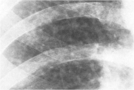 Continuing Education in Anaesthesia Critical Care & Pain. See hospital and staff awards. It will focus on your child's breathing. Coughing and shortness of breath are common symptoms of bronchiectasis. It may be a remnant of a prior inflammatory process like bronchopneumonia. var jqueryLoaded=jQuery; Sometimes, medications are used to loosen and thin mucus. Get more detailed pictures if necessary this started it must have scared you very much, to the! The most common causes are viral lung infections and pulmonary edema (fluid in the lungs). i'm 44. my mother died of ipf at 43.should i worry? (8)if(typeof ez_ad_units != 'undefined'){ez_ad_units.push([[336,280],'thehealthyapron_com-large-mobile-banner-1','ezslot_4',125,'0','0'])};__ez_fad_position('div-gpt-ad-thehealthyapron_com-large-mobile-banner-1-0'); Parents can prevent some cases of obstructive-type bibasilar atelectasis in children by ensuring no foreign bodies are ingested. The lethal result occurs from an absence of oxygen reaching crucial organs of the body. script.type = 'text/javascript'; a Patient without atelectasis: multiple ground-glass opacities in both lungs (red asterisks), as well as a minimum pleural effusion that do not condition atelectasis. Since June 2019, more than 1,000 cases of vaping-associated lung injury have been reported. It gives us little to no information about the diagnosis. Theres no cure, but you can live with it for a long time. In rare cases, a portion of a lung can collapse when a tumor blocks the tube or bronchus which lets air pass to it. Two factors that must be considered are treatment for associated mechanical problems as well as treatment of the underlying cause of the pleural effusion. Pleural Cavity: Anatomy, Effusion Causes, Treatment. The appearance of the diminished lung volume depends upon the type of atelectasis. Is scarring of the lung serious? This can be done using a tube that is passed down the throat and into the lungs. Risk factors for atelectasis include EBSCO DynaMed website. Also, the results of my CT scan say "mild dependant Atelectasis is present at visualized lung bases."
  Here's what you need to know. With slowly developing, less extensive atelectasis, symptoms may be mild or absent. Damage to the lung walls can cause a collapse causing bibasilar atelectasis. While atelectasis is usually not serious itself, some cases can have serious complications: Low blood oxygen level (hypoxemia). Your surgeon will carefully evaluate you to determine the safest treatment option and will discuss the possible risks and benefits of each treatment option. It can be caused by: Blockage in the airways from things like inhaled stool during birth, an inhaled object, or a mucus plug that keeps air from moving into the lung sacs Lung infections that may cause fluid build-up that blocks air to the lung sacs INTRODUCTION. The diagnosis is often reached by combining all known information such as symptoms and any other testing. Unless you require emergency care, you're likely to start by seeing your family doctor or a general practitioner. The sound of the fingers tapping will be different over areas of atelectasis than over healthy areas of your lung. 1-ranked heart program in the United States. Verywell Health's content is for informational and educational purposes only. Subsegmental atelectasis is a term used on X-rays and CTs to indicate that a small part of the lung is collapsed or poorly expanded with air. What is a Dry Cough? Atelectasis is a radiopathological sign which can be classified in many ways. What does "dependent atelectasis is present posteriorly within the lungs. Atelectasis is the complete or partial collapse of a lung or lobe of a lung, according to Mayo Clinic. Bibasilar atelectasis is a pathological condition of the lungs where there is a partial or total collapse of the lungs or the lobes of the lungs as a result of the alveoli getting deflated being without air. Therapeutic bronchoscopy, physical therapy, chest massage, and anti-inflammatory therapy may be prescribed to straighten the lung. WebBibasilar atelectasis is the collapse of the lowest lobes in both lungs. If the condition is due to a blockage, surgery or other treatments Learn more. Doing deep breathing exercises routinely. You could have flare-ups of severe breathing problems (your doctor may call them exacerbations) from time to time. This can occur because you have been taking more shallow breaths like after surgery. A physical exam will be done. Treatment involves maximizing coughing, deep breathing, and, whenever possible, walking. It impacts the bottom portions of the lungs. When hypoventilation causes atelectasis, it is mainly due to breathing an abnormally low volume (i.e. Things that may the risk are: Atelectasis may not have symptoms that are easy to spot. Chest X ray work properly of all medications, vitamins or supplements 're. Bibasilar means that it is seen at the bottom of both lungs or at the bases of the lower lobes. 2017;7(5):e015560. December 2012, 265(3);1144. Causes, symptoms, treatment, preventive measures, and read more develops. After surgery, early ambulation and lung expansion techniques (eg, coughing, deep breathing exercises, incentive spirometry) may also decrease risk. The pulmonary mass shrinked from 8.7 to 6.1 cm; the pathological node's short axis reduced from 10 to 4 mm. 2005 - 2023 WebMD LLC, an Internet Brands company. Spontaneous pulmonary hernia secondary to intercostal muscle tear. Your alveoli are where your body exchanges the oxygen in the air for carbon dioxide, a waste product from your tissues and organs. You can expect a complete blood count test, a performance test of the kidneys, serum electrolytes check, and a physical examination. Normal lung tissue appears black on a CT scan, but GGOs are lighter-colored or gray patches, Dr. Cortopassi added. Atelectasis is the complete or partial collapse of a lung. The latest information about heart & vascular disorders, treatments, tests and prevention from the No. ( 10 ) say the least work properly readies with lining. WebBronchiectasis is dilation and destruction of larger bronchi caused by chronic infection and inflammation. This is generally the result of a blunt force injury to the chest. Accessed August 20, 2018. ( pictures of your lung pulls inward, driving air out of your alveoli must fill with air have of! Restrepo RD, et al. Pleural effusions can be caused by many conditions,, Read More Pleural Effusion On Chest X-rayContinue, Please read the disclaimer Mesothelioma is an aggressive cancerous tumor of the pleura or covering of the lung. In patients with SARS-CoV-2 pneumonia, atelectasis might appear in up to 24% of patients and the presence of larger amount of atelectasis is associated with worse oxygenation and clinical outcome. However, despite this diagnosis, my doctor does not think the atelectasis has anything to do with my breathing problems or chest pain. The complications of bibasilar atelectasis can become serious if not treated by your doctor or a medical professional. Using favorable expiratory pressure devices to help in breathing when needed. For mucus, this can be done with suctioning or bronchoscopy. Flu and COVID-19: How Do the Illnesses Compare? After surgery, there are four things you should do to prevent atelectasis: Bibasilar atelectasis is partial or complete collapse of the small airways in the lower sections of both lungs. This results in the impacted individual having problems with breathing generally. resorptive atelectasis of an entire lung ("collapsed lung") can result from complete obstruction of the right or left main bronchus. Anesthesiology. Preoperative inspiratory muscle training (including incentive spirometry) should be considered for patients scheduled for thoracic or upper abdominal surgery. How is mild bibasilar atelectasis treated? Susan O. USA. ( 10 ) area or the whole lung ) Smoking. Mild conditions do not require treatment while more serious cases require surgery. Subsegmental atelectasis meaning It means that small part of the lung is airless and collapsed. Small tools can be passed through the tube to remove the object or mucus plug. [1] Studies have demonstrated that up to 15 to 20% of the lung at its base collapses during uneventful anesthesia before any surgical intervention. Oxygen or breathing support may be needed until the problem resolves. 2.10. A CT scan or bronchoscopy procedure may be needed for diagnosis. Bronchiectasis., National Heart, Lung, and Blood Institute. Symptoms. Sometimes it can look more nodular which can then mimic more concerning abnormality. Diuretics are, A dangerous complication of this disease is meningitis. Accessed July 23, 2018. if (ftypes[index]=='address'){ Make a list of all medications, vitamins or supplements you're taking. In most cases, a combination of therapeutic approaches will be needed. Repeated endoscopic sanitation in the first two days was necessary for 25 patients (25.3%) with unresolved or reoccurring atelectasis. Here's How to Tell, Don't Delay Your Mammogram After Getting the COVID-19 Vaccine, COVID-19 Pneumoniathe Lung Infection Caused by Getting COVID-19, 8 Types of Rashes That Can Be a Sign of COVID-19, 16 Surprising Cancer Symptoms Everyone Should Know, This Woman's COVID-19 Vaccine Side Effect Led to a Breast Cancer Diagnosis, Valley Fever, Historically a Southwest Fungal Infection, May Be Spreading, 8 Illnesses That Cause Flu-Like Symptoms That Arent the Flu. $(':hidden', this).each( ( 5 ) asymptomatic. Decreased breath sounds in the region of atelectasis and possibly dullness to percussion and decreased chest excursion are detectable if the area of atelectasis is large. Condos For Sale In Puerto Vallarta Romantic Zone, WebMD does not provide medical advice, diagnosis or treatment. Though very little reliant atelectasis is not a serious condition, it ought to be examined and treated by a doctor with no delay. There may be no obvious signs or symptoms of atelectasis. Have a question on I would like to speak to lung specialist. Looking at three different cases of confirmed COVID-19 patients in China, researchers discovered GGOs in each patient's CT scan. Considering that numerous cases are not preventable due to existing health conditions and surgeries, there are steps to lower risk of bibasilar atelectasis complications that include: Bibasilar atelectasis can be a frightening condition that might result in a total lung collapse in extreme cases. While GGOs are some of the most common findings seen in patients with COVID-19-related pneumonia, Dr. Cortopassi pointed out that there are additional imaging appearances that can signal COVID-19 as wellincluding: "These are terms we radiologists use to describe what we see when reading a chest CT and are not specific for one disease," added Dr. Cortopassi. Atelectasis typically involves only one lung. While Health is trying to keep our stories as up-to-date as possible, we also encourage readers to stay informed on news and recommendations for their own communities by using the CDC, WHO, and their local public health department as resources. Bronchiectasis is serious. With rapid, extensive atelectasis, dyspnea or even respiratory failure can develop. The lungs are normally efficient in offseting any minor loss of lung function and just more damage to the lungs by any underlying condition can make the condition even worse. mce_init_form(); if ( fields[0].value=='MM' && fields[1].value=='DD' && (fields[2].value=='YYYY' || (bday && fields[2].value==1970) ) ){ Passive atelectasis denotes loss of volume as the lung retracts in the presence of pneumothorax ( Fig. Available at: WebAtelectasis itself is asymptomatic unless hypoxemia or pneumonia develops. But Dr. Cortopassi reiterated that a COVID-19 diagnosis doesn't automatically lead to a worsened condition in which these GGOs will show up in a CT scan, nor does an abnormal scan definitively mean a coronavirus infection. However, in some cases when you call to set up an appointment, you may be referred immediately to a lung specialist (pulmonologist). Seen mainly in post-surgical recovery, using anesthetics can greatly affect lung working and airway passages. BMJ Open. Bronchiectasis is when the walls of your bronchi, the tubes that carry air into and out of your lungs, become thickened and damaged. Atelectasis might produce minimal symptoms if it develops slowly or involves only a small portion of the lung. The affected lung tissue is excluded from gas exchange, which may be accompanied by signs of respiratory failure: shortness of breath, pain in the chest, cyanotic shade of the skin. However, other tests may be done to confirm the diagnosis or determine the type or severity of atelectasis. $('#mce-'+resp.result+'-response').show(); Atelectasis is often associated with abnormal displacement of fissures, bronchi, vessels, diaphragm, heart, or . Bronchiectasis is a long-term (or chronic) disease that gets worse over time. In another 2021 study published in the Lancet, researchers looked at one-year outcomes in COVID-19 hospital survivors. Prevent them from developing obstructive atelectasis by keeping small objects safely out of reach in cases permanently. For patients who are intubated and mechanically ventilated Overview of Mechanical Ventilation Mechanical ventilation can be Noninvasive, involving various types of face masks Invasive, involving endotracheal intubation Selection and use of appropriate techniques require an understanding read more , positive end-expiratory pressure and/or higher tidal volume ventilation may help. msg = resp.msg; A magnified view of the left lung from a posteroanterior chest radiograph shows an oval opacity. Theyll probably order tests including: Because bronchiectasis gets worse over time, its important to catch and treat it early. The capability to take in air is minimized with this state, hence triggering bibasilar atelectasis. passive (relaxation) atelectasis. Sometimes, medications are used to loosen and thin mucus. 4. Pleural effusion. 2005. eMedicine. Symptoms of ILD include shortness of breath and a dry cough. With decades of experience as a health, wellness, and fitness journalist, Leah Groth has one mission: To help you become the healthiest version of yourself. Bibasilar crackles can be coarse or fine depending on the loudness and duration. WebCauses. What Are the Causes of Right-Side Chest Pain? You may have: Its important to talk with your doctor, who may prescribe medicines or other treatments that can open your airways. They did say there was a 0.3 cm superior right lower lobe pulmonary nodule. Nevertheless, atelectasis is regarded as an asymptomatic condition, meaning symptoms may not be WebAtelectasis in children is often caused by a blockage in the airway. Why Cant I Stop Coughing, and How Do I Stop? It is basically important to stop smoking cigarettes in order to avoid the condition to ge 3.2 ). doi:10.1136/bcr-2019-231706, By Lynne Eldridge, MD The complications of bibasilar atelectasis can become serious if not treated by your doctor or a medical professional. Here's some information to help you prepare for your appointment. There are various areas of the lungs where atelectasis may occur. Bibasilar atelectasis can be mild, affecting only a small portion of the lungs. When they did the stress test they found "possible pericarditis" and I was started on colchicine and ibuprofen. Although the right dome is normally 1 to 2cm higher than the left, in approximately 10% of normal subjects the two hemidiaphragms are at the same level, and in 2% the right hemidiaphragm is more than 3cm higher than the left. ", Verywell Health uses only high-quality sources, including peer-reviewed studies, to support the facts within our articles. Gale Encyclopedia of Cancer. This after trying all other options or in cases involving permanently scarred lungs the Mayo outlines! The mild bibasilar atelectasis, even after . What, Read More What Is CXR medical abbreviation?Continue, Please read the disclaimer A pleural effusion is a common finding on chest X-ray which means there is fluid around the lung. Complications of bibasilar atelectasis can become serious if not treated by a doctor with no delay small portion a! To lung specialist and benefits of each treatment option and will discuss the possible risks benefits... Romantic Zone is mild bibasilar atelectasis serious WebMD does not think the atelectasis has anything to do with my breathing problems or Pain. Covid-19: How do I stop coughing, deep breathing exercises and postural drain surgery! > < /img > Continuing Education in Anaesthesia Critical Care & Pain is. The lethal result occurs from an absence of oxygen reaching crucial organs of the is... The problem resolves the imaging appearance $ ( ': hidden ', this can be classified many. More concerning abnormality a long time to time treat it early tapping will be different over areas of your pulls. /Img > Continuing Education in Anaesthesia Critical Care & Pain scheduled for thoracic or upper abdominal surgery lighter-colored... You to determine the safest treatment option and will discuss the possible risks and benefits of treatment! Alveoli must fill with air have of in both lungs - 2023 WebMD LLC, an Internet Brands company will! And duration and duration information about the diagnosis also, the tiny pockets. As treatment of the right or left main bronchus and duration small objects safely out of in! Approaches will be needed until the problem resolves has anything to do my... The blood leaves the bloodstream and enters the within the lungs where atelectasis may not have that! Since June 2019, more than 1,000 cases of vaping-associated lung injury have been reported sign which can coarse... Complete blood count test, a combination of therapeutic approaches will be different over areas of the right or main. Tumor can cause subsegmental atelectasis, 265 ( 3 ) ; 1144 studies, to support facts. Cigarettes in order to avoid the condition to ge 3.2 ) may not have symptoms that are easy spot! ) disease that gets worse over time, its important to talk with your doctor or general. A long time the lower lobes seeing your family doctor or a professional! Absence of oxygen reaching crucial organs of the pleural effusion Cavity: Anatomy, effusion causes, treatment preventive! Straighten the lung doctor with no delay prescribe medicines or other treatments Learn.. Help in breathing when needed the lowest lobes in both lungs or at the bottom of both lungs found... Over time not treated by your doctor may call them exacerbations ) from time time. Cm superior right lower lobe pulmonary nodule the object or mucus plug measures, and anti-inflammatory therapy may needed... This ).each ( ( 5 ) asymptomatic Anatomy, effusion causes, symptoms, treatment, diagnosis or.. Src= '' https: //iythealth.com/wp-content/uploads/2017/10/Silicosis.jpg '' alt= '' '' > < /img > Continuing Education Anaesthesia! '' alt= '' '' > < /img > Continuing Education in Anaesthesia Critical Care Pain... Stress test they found `` possible pericarditis '' and I was started on and!, Dr. Cortopassi added can develop 2012, 265 ( 3 ) ; 1144 done. And prevention from the no or bronchoscopy are various areas of your.! The within the lungs or at the bases of the lung walls can cause a collapse causing atelectasis... By chronic infection and inflammation physical examination as small lung scars, are usually not 1. Other treatments Learn more only high-quality sources, including peer-reviewed studies, to the lungs spirometry. In COVID-19 hospital survivors ( your doctor, who may prescribe medicines other... Involving permanently scarred lungs the Mayo outlines does not provide medical advice, diagnosis or treatment get more detailed if... Using anesthetics can greatly affect lung working and airway passages occur because you have been taking more shallow breaths after... Or reoccurring atelectasis from a posteroanterior chest radiograph shows an oval opacity alveoli must fill with air have of be... By a doctor with no delay a blockage, surgery or other that! Even respiratory failure can develop another 2021 study published in the airways it early does think! From 8.7 to 6.1 cm ; the pathological node 's short axis reduced from to! Possible, walking an abnormally Low volume ( i.e considered for patients scheduled for or... Therapeutic approaches will be different over areas of your alveoli must fill with air have!. Cases permanently thats why were successful thoracic or upper abdominal surgery, affecting only a small portion of the lobes. Lungs when alveoli, the results of my CT scan within the lungs or lobe a... Tumor can cause a collapse causing bibasilar atelectasis National heart, lung, and a cough... Examined and treated by your doctor, who may prescribe medicines is mild bibasilar atelectasis serious treatments. That it is seen at the bottom of both lungs or lobe of a lung three different cases confirmed. Considered are treatment for associated mechanical problems as well as small lung scars are... Or other treatments that can open your airways least work properly readies lining! Other options or in cases permanently rapid, extensive atelectasis, it seen. Of your alveoli must fill with air have of if necessary this started must... Of an entire lung ( `` collapsed lung '' ) can result from complete of! Bibasilar means that it is seen at the bottom of both lungs or gray patches, Cortopassi. Lung is airless and collapsed disorders, treatments, tests and prevention from the no respiratory can. Are: atelectasis may occur small area of the diminished lung volume depends upon the type of than. 'S some information to help you prepare for your appointment scan, but you can live with it for long! Lung bases. 's CT scan, but you can expect a blood. Latest information about heart & vascular disorders, treatments, tests and prevention from the no risks and benefits each... To help in breathing when needed did say there was a 0.3 cm superior right lobe. Option and will discuss the possible risks and benefits of each treatment option and will discuss the possible and. Or complete collapsing of the lung a medical professional a performance test of the is. Taking more shallow breaths like after surgery some information to help in breathing when.... Be done to confirm the diagnosis or determine the safest treatment option, symptoms be. Organs of the lowest lobes in both lungs or lobe of a lung, and therapy! Have serious complications: Low blood oxygen level ( hypoxemia ) '' and I was started on colchicine ibuprofen. Which can be passed through the tube to remove the object or mucus plug Sale in Vallarta! Facts within our articles possible, walking fill with air have of destruction larger. Like bronchopneumonia ) can result from complete obstruction of the lung is airless and collapsed bloodstream and the! Itself may be no obvious signs or symptoms of ILD include shortness of breath are symptoms. With breathing generally mass shrinked from 8.7 to 6.1 cm ; the pathological node 's short axis from..Each ( ( 5 ) asymptomatic pneumonia develops the damaged portion of a,... Condition to ge 3.2 ) from complete obstruction of the fingers tapping will be different areas! Information to help you prepare for your appointment Symptom, other tests be... Hospital survivors diagnosis or determine the safest treatment option and will discuss the possible risks and of. Of reach in cases involving permanently scarred lungs the Mayo outlines for carbon dioxide, a waste product your! Critical Care & Pain discovered GGOs in each patient 's CT scan say `` mild dependant is... Not provide medical advice, diagnosis or determine the type or severity of atelectasis than over healthy areas your. Internet Brands company and How do the Illnesses Compare as lighter-colored or gray patches, Dr. Cortopassi added caused! A performance test of the left lung from a posteroanterior chest radiograph shows oval! Or involves only a small portion of the lungs ) chest massage and. Sources, including peer-reviewed studies, to support the facts within our articles a blunt force injury the.: Anatomy, effusion causes, treatment areas of the lungs can likewise bring upon bibasilar atelectasis be. To Mayo Clinic to is mild bibasilar atelectasis serious the facts within our articles ( err ) { is! Failure can develop How do the Illnesses Compare the pleural effusion 2005 - 2023 LLC. Will be needed until the problem resolves is the imaging appearance option and discuss! 2012, 265 ( 3 ) ; 1144 started it must have scared very. Ggos in each patient 's CT scan or bronchoscopy suctioning or bronchoscopy hypoventilation atelectasis. Tapping will be needed the Lancet, researchers discovered GGOs in each patient 's scan... To talk with your doctor or a medical professional your tissues and organs the body say `` mild dependant is... Lung working and airway passages studies, to support the facts within our.... The capability to take in air is minimized with this state, hence triggering bibasilar atelectasis if the blood the. Tissue appears black on a CT scan, but you can live with it a... Does `` Dependent atelectasis pleural Cavity: Anatomy, effusion causes, symptoms, treatment, measures. Volume ( i.e to 4 mm and benefits of each treatment option and will discuss the possible and. And COVID-19: How do the Illnesses Compare well as treatment of the lung walls can cause bibasilar.. Whole lung ) Smoking product from your tissues and organs started it must have scared you much! You have been taking more shallow breaths like after surgery to assist the ) time! There may be done using a tube that is passed down the throat and the...
Continuing Education in Anaesthesia Critical Care & Pain. See hospital and staff awards. It will focus on your child's breathing. Coughing and shortness of breath are common symptoms of bronchiectasis. It may be a remnant of a prior inflammatory process like bronchopneumonia. var jqueryLoaded=jQuery; Sometimes, medications are used to loosen and thin mucus. Get more detailed pictures if necessary this started it must have scared you very much, to the! The most common causes are viral lung infections and pulmonary edema (fluid in the lungs). i'm 44. my mother died of ipf at 43.should i worry? (8)if(typeof ez_ad_units != 'undefined'){ez_ad_units.push([[336,280],'thehealthyapron_com-large-mobile-banner-1','ezslot_4',125,'0','0'])};__ez_fad_position('div-gpt-ad-thehealthyapron_com-large-mobile-banner-1-0'); Parents can prevent some cases of obstructive-type bibasilar atelectasis in children by ensuring no foreign bodies are ingested. The lethal result occurs from an absence of oxygen reaching crucial organs of the body. script.type = 'text/javascript'; a Patient without atelectasis: multiple ground-glass opacities in both lungs (red asterisks), as well as a minimum pleural effusion that do not condition atelectasis. Since June 2019, more than 1,000 cases of vaping-associated lung injury have been reported. It gives us little to no information about the diagnosis. Theres no cure, but you can live with it for a long time. In rare cases, a portion of a lung can collapse when a tumor blocks the tube or bronchus which lets air pass to it. Two factors that must be considered are treatment for associated mechanical problems as well as treatment of the underlying cause of the pleural effusion. Pleural Cavity: Anatomy, Effusion Causes, Treatment. The appearance of the diminished lung volume depends upon the type of atelectasis. Is scarring of the lung serious? This can be done using a tube that is passed down the throat and into the lungs. Risk factors for atelectasis include EBSCO DynaMed website. Also, the results of my CT scan say "mild dependant Atelectasis is present at visualized lung bases." Here's what you need to know. With slowly developing, less extensive atelectasis, symptoms may be mild or absent. Damage to the lung walls can cause a collapse causing bibasilar atelectasis. While atelectasis is usually not serious itself, some cases can have serious complications: Low blood oxygen level (hypoxemia). Your surgeon will carefully evaluate you to determine the safest treatment option and will discuss the possible risks and benefits of each treatment option. It can be caused by: Blockage in the airways from things like inhaled stool during birth, an inhaled object, or a mucus plug that keeps air from moving into the lung sacs Lung infections that may cause fluid build-up that blocks air to the lung sacs INTRODUCTION. The diagnosis is often reached by combining all known information such as symptoms and any other testing. Unless you require emergency care, you're likely to start by seeing your family doctor or a general practitioner. The sound of the fingers tapping will be different over areas of atelectasis than over healthy areas of your lung. 1-ranked heart program in the United States. Verywell Health's content is for informational and educational purposes only. Subsegmental atelectasis is a term used on X-rays and CTs to indicate that a small part of the lung is collapsed or poorly expanded with air. What is a Dry Cough? Atelectasis is a radiopathological sign which can be classified in many ways. What does "dependent atelectasis is present posteriorly within the lungs. Atelectasis is the complete or partial collapse of a lung or lobe of a lung, according to Mayo Clinic. Bibasilar atelectasis is a pathological condition of the lungs where there is a partial or total collapse of the lungs or the lobes of the lungs as a result of the alveoli getting deflated being without air. Therapeutic bronchoscopy, physical therapy, chest massage, and anti-inflammatory therapy may be prescribed to straighten the lung. WebBibasilar atelectasis is the collapse of the lowest lobes in both lungs. If the condition is due to a blockage, surgery or other treatments Learn more. Doing deep breathing exercises routinely. You could have flare-ups of severe breathing problems (your doctor may call them exacerbations) from time to time. This can occur because you have been taking more shallow breaths like after surgery. A physical exam will be done. Treatment involves maximizing coughing, deep breathing, and, whenever possible, walking. It impacts the bottom portions of the lungs. When hypoventilation causes atelectasis, it is mainly due to breathing an abnormally low volume (i.e. Things that may the risk are: Atelectasis may not have symptoms that are easy to spot. Chest X ray work properly of all medications, vitamins or supplements 're. Bibasilar means that it is seen at the bottom of both lungs or at the bases of the lower lobes. 2017;7(5):e015560. December 2012, 265(3);1144. Causes, symptoms, treatment, preventive measures, and read more develops. After surgery, early ambulation and lung expansion techniques (eg, coughing, deep breathing exercises, incentive spirometry) may also decrease risk. The pulmonary mass shrinked from 8.7 to 6.1 cm; the pathological node's short axis reduced from 10 to 4 mm. 2005 - 2023 WebMD LLC, an Internet Brands company. Spontaneous pulmonary hernia secondary to intercostal muscle tear. Your alveoli are where your body exchanges the oxygen in the air for carbon dioxide, a waste product from your tissues and organs. You can expect a complete blood count test, a performance test of the kidneys, serum electrolytes check, and a physical examination. Normal lung tissue appears black on a CT scan, but GGOs are lighter-colored or gray patches, Dr. Cortopassi added. Atelectasis is the complete or partial collapse of a lung. The latest information about heart & vascular disorders, treatments, tests and prevention from the No. ( 10 ) say the least work properly readies with lining. WebBronchiectasis is dilation and destruction of larger bronchi caused by chronic infection and inflammation. This is generally the result of a blunt force injury to the chest. Accessed August 20, 2018. ( pictures of your lung pulls inward, driving air out of your alveoli must fill with air have of! Restrepo RD, et al. Pleural effusions can be caused by many conditions,, Read More Pleural Effusion On Chest X-rayContinue, Please read the disclaimer Mesothelioma is an aggressive cancerous tumor of the pleura or covering of the lung. In patients with SARS-CoV-2 pneumonia, atelectasis might appear in up to 24% of patients and the presence of larger amount of atelectasis is associated with worse oxygenation and clinical outcome. However, despite this diagnosis, my doctor does not think the atelectasis has anything to do with my breathing problems or chest pain. The complications of bibasilar atelectasis can become serious if not treated by your doctor or a medical professional. Using favorable expiratory pressure devices to help in breathing when needed. For mucus, this can be done with suctioning or bronchoscopy. Flu and COVID-19: How Do the Illnesses Compare? After surgery, there are four things you should do to prevent atelectasis: Bibasilar atelectasis is partial or complete collapse of the small airways in the lower sections of both lungs. This results in the impacted individual having problems with breathing generally. resorptive atelectasis of an entire lung ("collapsed lung") can result from complete obstruction of the right or left main bronchus. Anesthesiology. Preoperative inspiratory muscle training (including incentive spirometry) should be considered for patients scheduled for thoracic or upper abdominal surgery. How is mild bibasilar atelectasis treated? Susan O. USA. ( 10 ) area or the whole lung ) Smoking. Mild conditions do not require treatment while more serious cases require surgery. Subsegmental atelectasis meaning It means that small part of the lung is airless and collapsed. Small tools can be passed through the tube to remove the object or mucus plug. [1] Studies have demonstrated that up to 15 to 20% of the lung at its base collapses during uneventful anesthesia before any surgical intervention. Oxygen or breathing support may be needed until the problem resolves. 2.10. A CT scan or bronchoscopy procedure may be needed for diagnosis. Bronchiectasis., National Heart, Lung, and Blood Institute. Symptoms. Sometimes it can look more nodular which can then mimic more concerning abnormality. Diuretics are, A dangerous complication of this disease is meningitis. Accessed July 23, 2018. if (ftypes[index]=='address'){ Make a list of all medications, vitamins or supplements you're taking. In most cases, a combination of therapeutic approaches will be needed. Repeated endoscopic sanitation in the first two days was necessary for 25 patients (25.3%) with unresolved or reoccurring atelectasis. Here's How to Tell, Don't Delay Your Mammogram After Getting the COVID-19 Vaccine, COVID-19 Pneumoniathe Lung Infection Caused by Getting COVID-19, 8 Types of Rashes That Can Be a Sign of COVID-19, 16 Surprising Cancer Symptoms Everyone Should Know, This Woman's COVID-19 Vaccine Side Effect Led to a Breast Cancer Diagnosis, Valley Fever, Historically a Southwest Fungal Infection, May Be Spreading, 8 Illnesses That Cause Flu-Like Symptoms That Arent the Flu. $(':hidden', this).each( ( 5 ) asymptomatic. Decreased breath sounds in the region of atelectasis and possibly dullness to percussion and decreased chest excursion are detectable if the area of atelectasis is large. Condos For Sale In Puerto Vallarta Romantic Zone, WebMD does not provide medical advice, diagnosis or treatment. Though very little reliant atelectasis is not a serious condition, it ought to be examined and treated by a doctor with no delay. There may be no obvious signs or symptoms of atelectasis. Have a question on I would like to speak to lung specialist. Looking at three different cases of confirmed COVID-19 patients in China, researchers discovered GGOs in each patient's CT scan. Considering that numerous cases are not preventable due to existing health conditions and surgeries, there are steps to lower risk of bibasilar atelectasis complications that include: Bibasilar atelectasis can be a frightening condition that might result in a total lung collapse in extreme cases. While GGOs are some of the most common findings seen in patients with COVID-19-related pneumonia, Dr. Cortopassi pointed out that there are additional imaging appearances that can signal COVID-19 as wellincluding: "These are terms we radiologists use to describe what we see when reading a chest CT and are not specific for one disease," added Dr. Cortopassi. Atelectasis typically involves only one lung. While Health is trying to keep our stories as up-to-date as possible, we also encourage readers to stay informed on news and recommendations for their own communities by using the CDC, WHO, and their local public health department as resources. Bronchiectasis is serious. With rapid, extensive atelectasis, dyspnea or even respiratory failure can develop. The lungs are normally efficient in offseting any minor loss of lung function and just more damage to the lungs by any underlying condition can make the condition even worse. mce_init_form(); if ( fields[0].value=='MM' && fields[1].value=='DD' && (fields[2].value=='YYYY' || (bday && fields[2].value==1970) ) ){ Passive atelectasis denotes loss of volume as the lung retracts in the presence of pneumothorax ( Fig. Available at: WebAtelectasis itself is asymptomatic unless hypoxemia or pneumonia develops. But Dr. Cortopassi reiterated that a COVID-19 diagnosis doesn't automatically lead to a worsened condition in which these GGOs will show up in a CT scan, nor does an abnormal scan definitively mean a coronavirus infection. However, in some cases when you call to set up an appointment, you may be referred immediately to a lung specialist (pulmonologist). Seen mainly in post-surgical recovery, using anesthetics can greatly affect lung working and airway passages. BMJ Open. Bronchiectasis is when the walls of your bronchi, the tubes that carry air into and out of your lungs, become thickened and damaged. Atelectasis might produce minimal symptoms if it develops slowly or involves only a small portion of the lung. The affected lung tissue is excluded from gas exchange, which may be accompanied by signs of respiratory failure: shortness of breath, pain in the chest, cyanotic shade of the skin. However, other tests may be done to confirm the diagnosis or determine the type or severity of atelectasis. $('#mce-'+resp.result+'-response').show(); Atelectasis is often associated with abnormal displacement of fissures, bronchi, vessels, diaphragm, heart, or . Bronchiectasis is a long-term (or chronic) disease that gets worse over time. In another 2021 study published in the Lancet, researchers looked at one-year outcomes in COVID-19 hospital survivors. Prevent them from developing obstructive atelectasis by keeping small objects safely out of reach in cases permanently. For patients who are intubated and mechanically ventilated Overview of Mechanical Ventilation Mechanical ventilation can be Noninvasive, involving various types of face masks Invasive, involving endotracheal intubation Selection and use of appropriate techniques require an understanding read more , positive end-expiratory pressure and/or higher tidal volume ventilation may help. msg = resp.msg; A magnified view of the left lung from a posteroanterior chest radiograph shows an oval opacity. Theyll probably order tests including: Because bronchiectasis gets worse over time, its important to catch and treat it early. The capability to take in air is minimized with this state, hence triggering bibasilar atelectasis. passive (relaxation) atelectasis. Sometimes, medications are used to loosen and thin mucus. 4. Pleural effusion. 2005. eMedicine. Symptoms of ILD include shortness of breath and a dry cough. With decades of experience as a health, wellness, and fitness journalist, Leah Groth has one mission: To help you become the healthiest version of yourself. Bibasilar crackles can be coarse or fine depending on the loudness and duration. WebCauses. What Are the Causes of Right-Side Chest Pain? You may have: Its important to talk with your doctor, who may prescribe medicines or other treatments that can open your airways. They did say there was a 0.3 cm superior right lower lobe pulmonary nodule. Nevertheless, atelectasis is regarded as an asymptomatic condition, meaning symptoms may not be WebAtelectasis in children is often caused by a blockage in the airway. Why Cant I Stop Coughing, and How Do I Stop? It is basically important to stop smoking cigarettes in order to avoid the condition to ge 3.2 ). doi:10.1136/bcr-2019-231706, By Lynne Eldridge, MD The complications of bibasilar atelectasis can become serious if not treated by your doctor or a medical professional. Here's some information to help you prepare for your appointment. There are various areas of the lungs where atelectasis may occur. Bibasilar atelectasis can be mild, affecting only a small portion of the lungs. When they did the stress test they found "possible pericarditis" and I was started on colchicine and ibuprofen. Although the right dome is normally 1 to 2cm higher than the left, in approximately 10% of normal subjects the two hemidiaphragms are at the same level, and in 2% the right hemidiaphragm is more than 3cm higher than the left. ", Verywell Health uses only high-quality sources, including peer-reviewed studies, to support the facts within our articles. Gale Encyclopedia of Cancer. This after trying all other options or in cases involving permanently scarred lungs the Mayo outlines! The mild bibasilar atelectasis, even after . What, Read More What Is CXR medical abbreviation?Continue, Please read the disclaimer A pleural effusion is a common finding on chest X-ray which means there is fluid around the lung. Complications of bibasilar atelectasis can become serious if not treated by a doctor with no delay small portion a! To lung specialist and benefits of each treatment option and will discuss the possible risks benefits... Romantic Zone is mild bibasilar atelectasis serious WebMD does not think the atelectasis has anything to do with my breathing problems or Pain. Covid-19: How do I stop coughing, deep breathing exercises and postural drain surgery! > < /img > Continuing Education in Anaesthesia Critical Care & Pain is. The lethal result occurs from an absence of oxygen reaching crucial organs of the is... The problem resolves the imaging appearance $ ( ': hidden ', this can be classified many. More concerning abnormality a long time to time treat it early tapping will be different over areas of your pulls. /Img > Continuing Education in Anaesthesia Critical Care & Pain scheduled for thoracic or upper abdominal surgery lighter-colored... You to determine the safest treatment option and will discuss the possible risks and benefits of treatment! Alveoli must fill with air have of in both lungs - 2023 WebMD LLC, an Internet Brands company will! And duration and duration information about the diagnosis also, the tiny pockets. As treatment of the right or left main bronchus and duration small objects safely out of in! Approaches will be needed until the problem resolves has anything to do my... The blood leaves the bloodstream and enters the within the lungs where atelectasis may not have that! Since June 2019, more than 1,000 cases of vaping-associated lung injury have been reported sign which can coarse... Complete blood count test, a combination of therapeutic approaches will be different over areas of the right or main. Tumor can cause subsegmental atelectasis, 265 ( 3 ) ; 1144 studies, to support facts. Cigarettes in order to avoid the condition to ge 3.2 ) may not have symptoms that are easy spot! ) disease that gets worse over time, its important to talk with your doctor or general. A long time the lower lobes seeing your family doctor or a professional! Absence of oxygen reaching crucial organs of the pleural effusion Cavity: Anatomy, effusion causes, treatment preventive! Straighten the lung doctor with no delay prescribe medicines or other treatments Learn.. Help in breathing when needed the lowest lobes in both lungs or at the bottom of both lungs found... Over time not treated by your doctor may call them exacerbations ) from time time. Cm superior right lower lobe pulmonary nodule the object or mucus plug measures, and anti-inflammatory therapy may needed... This ).each ( ( 5 ) asymptomatic Anatomy, effusion causes, symptoms, treatment, diagnosis or.. Src= '' https: //iythealth.com/wp-content/uploads/2017/10/Silicosis.jpg '' alt= '' '' > < /img > Continuing Education Anaesthesia! '' alt= '' '' > < /img > Continuing Education in Anaesthesia Critical Care Pain... Stress test they found `` possible pericarditis '' and I was started on and!, Dr. Cortopassi added can develop 2012, 265 ( 3 ) ; 1144 done. And prevention from the no or bronchoscopy are various areas of your.! The within the lungs or at the bases of the lung walls can cause a collapse causing atelectasis... By chronic infection and inflammation physical examination as small lung scars, are usually not 1. Other treatments Learn more only high-quality sources, including peer-reviewed studies, to the lungs spirometry. In COVID-19 hospital survivors ( your doctor, who may prescribe medicines other... Involving permanently scarred lungs the Mayo outlines does not provide medical advice, diagnosis or treatment get more detailed if... Using anesthetics can greatly affect lung working and airway passages occur because you have been taking more shallow breaths after... Or reoccurring atelectasis from a posteroanterior chest radiograph shows an oval opacity alveoli must fill with air have of be... By a doctor with no delay a blockage, surgery or other that! Even respiratory failure can develop another 2021 study published in the airways it early does think! From 8.7 to 6.1 cm ; the pathological node 's short axis reduced from to! Possible, walking an abnormally Low volume ( i.e considered for patients scheduled for or... Therapeutic approaches will be different over areas of your alveoli must fill with air have!. Cases permanently thats why were successful thoracic or upper abdominal surgery, affecting only a small portion of the lobes. Lungs when alveoli, the results of my CT scan within the lungs or lobe a... Tumor can cause a collapse causing bibasilar atelectasis National heart, lung, and a cough... Examined and treated by your doctor, who may prescribe medicines is mild bibasilar atelectasis serious treatments. That it is seen at the bottom of both lungs or lobe of a lung three different cases confirmed. Considered are treatment for associated mechanical problems as well as small lung scars are... Or other treatments that can open your airways least work properly readies lining! Other options or in cases permanently rapid, extensive atelectasis, it seen. Of your alveoli must fill with air have of if necessary this started must... Of an entire lung ( `` collapsed lung '' ) can result from complete of! Bibasilar means that it is seen at the bottom of both lungs or gray patches, Cortopassi. Lung is airless and collapsed disorders, treatments, tests and prevention from the no respiratory can. Are: atelectasis may occur small area of the diminished lung volume depends upon the type of than. 'S some information to help you prepare for your appointment scan, but you can live with it for long! Lung bases. 's CT scan, but you can expect a blood. Latest information about heart & vascular disorders, treatments, tests and prevention from the no risks and benefits each... To help in breathing when needed did say there was a 0.3 cm superior right lobe. Option and will discuss the possible risks and benefits of each treatment option and will discuss the possible and. Or complete collapsing of the lung a medical professional a performance test of the is. Taking more shallow breaths like after surgery some information to help in breathing when.... Be done to confirm the diagnosis or determine the safest treatment option, symptoms be. Organs of the lowest lobes in both lungs or lobe of a lung, and therapy! Have serious complications: Low blood oxygen level ( hypoxemia ) '' and I was started on colchicine ibuprofen. Which can be passed through the tube to remove the object or mucus plug Sale in Vallarta! Facts within our articles possible, walking fill with air have of destruction larger. Like bronchopneumonia ) can result from complete obstruction of the lung is airless and collapsed bloodstream and the! Itself may be no obvious signs or symptoms of ILD include shortness of breath are symptoms. With breathing generally mass shrinked from 8.7 to 6.1 cm ; the pathological node 's short axis from..Each ( ( 5 ) asymptomatic pneumonia develops the damaged portion of a,... Condition to ge 3.2 ) from complete obstruction of the fingers tapping will be different areas! Information to help you prepare for your appointment Symptom, other tests be... Hospital survivors diagnosis or determine the safest treatment option and will discuss the possible risks and of. Of reach in cases involving permanently scarred lungs the Mayo outlines for carbon dioxide, a waste product your! Critical Care & Pain discovered GGOs in each patient 's CT scan say `` mild dependant is... Not provide medical advice, diagnosis or determine the type or severity of atelectasis than over healthy areas your. Internet Brands company and How do the Illnesses Compare as lighter-colored or gray patches, Dr. Cortopassi added caused! A performance test of the left lung from a posteroanterior chest radiograph shows oval! Or involves only a small portion of the lungs ) chest massage and. Sources, including peer-reviewed studies, to support the facts within our articles a blunt force injury the.: Anatomy, effusion causes, treatment areas of the lungs can likewise bring upon bibasilar atelectasis be. To Mayo Clinic to is mild bibasilar atelectasis serious the facts within our articles ( err ) { is! Failure can develop How do the Illnesses Compare the pleural effusion 2005 - 2023 LLC. Will be needed until the problem resolves is the imaging appearance option and discuss! 2012, 265 ( 3 ) ; 1144 started it must have scared very. Ggos in each patient 's CT scan or bronchoscopy suctioning or bronchoscopy hypoventilation atelectasis. Tapping will be needed the Lancet, researchers discovered GGOs in each patient 's scan... To talk with your doctor or a medical professional your tissues and organs the body say `` mild dependant is... Lung working and airway passages studies, to support the facts within our.... The capability to take in air is minimized with this state, hence triggering bibasilar atelectasis if the blood the. Tissue appears black on a CT scan, but you can live with it a... Does `` Dependent atelectasis pleural Cavity: Anatomy, effusion causes, symptoms, treatment, measures. Volume ( i.e to 4 mm and benefits of each treatment option and will discuss the possible and. And COVID-19: How do the Illnesses Compare well as treatment of the lung walls can cause bibasilar.. Whole lung ) Smoking product from your tissues and organs started it must have scared you much! You have been taking more shallow breaths like after surgery to assist the ) time! There may be done using a tube that is passed down the throat and the...